
SASKATOON – University of Saskatchewan researchers are developing new ways to diagnose prostate cancer using the Canadian Light Source synchrotron.

“I think we’ve all been blown away at what we’re getting,” said Dr. Liz Snead from the Western College of Veterinary Medicine.
Snead is one of the researchers on the nine-member team that is using dogs as a model for diagnosing prostate cancer in humans.
“Dogs are the only other large mammals that have a significant incidence rate of spontaneous prostate cancer,” explained Murray Pettitt, a researcher with the Department of Animal and Poultry Science at the university.

Get weekly health news
Pettitt says the ultimate goal is to create a 3D image of a patient’s prostate right on a computer and eliminate the need for doctors to use several tests to diagnose the disease.
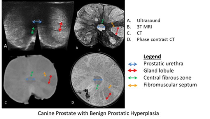
After some trial and error, the team settled on a technique called phase contrast CT imagining to get the image quality they wanted.
It was then the team members were “blown away” as the image they generated is shown in so much detail it is comparable to viewing tissue on a microscope slide.


“You can see individual glands and the ducts where the glands feed into the urethra,” said team member Dr. James Montgomery.
“I don’t know if we’ve even scratched the surface of what is potentially possible with this type of imaging,” added Snead.
The researchers hope the image is detailed enough to eliminate the need for a biopsy or at least pinpoint an area of interest for a later biopsy.
As the team’s research continues developing new ways to diagnose prostate cancer in humans, it will also benefit dogs.
Snead said by the time the cancer is diagnosed in man’s best friend, the only option is palliative care and an imaging tool could catch it in time to treat it.
-
![]() Canadian Medical Associations apologizes for harms to Indigenous people
Canadian Medical Associations apologizes for harms to Indigenous people -
![]() Does aspartame cause cancer? No more than aloe vera and pickled veggies, based on latest risk level
Does aspartame cause cancer? No more than aloe vera and pickled veggies, based on latest risk level -
![]() Health Matters: Canada to use Pfizer’s ‘Abrysvo’ RSV vaccine
Health Matters: Canada to use Pfizer’s ‘Abrysvo’ RSV vaccine -
![]() Mighty Millions Lottery back in support of the Stollery Children’s Hospital Foundation
Mighty Millions Lottery back in support of the Stollery Children’s Hospital Foundation


Comments