

Their skulls have ridges, are indented, and have white space where brain tissue should be. In a new case study, Brazilian and American doctors reveal a series of tragic scans that show Zika virus’ destruction on unborn babies’ development.

The photos, released by doctors out of the Federal University of Rio de Janeiro and Harvard Medical School, were put together to help radiologists take a closer look at the full scale of issues stemming from Zika virus.
“Microcephaly is just one of several radiological features,” Dr. Fernanda Tovar-Moll, the study’s lead author, said.
“Imaging is essential for identifying the presence and the severity of the structural changes induced by the infection, especially in the central nervous system,” Tover-Moll explained.
READ MORE: These are the tell-tale symptoms of Zika virus, according to a new case study
The medical community has already warned that Zika appears to be most dangerous when passed on from an expectant mom to her baby in the first trimester.
Health officials in El Salvador, Brazil, Jamaica, Ecuador, Honduras and Colombia told residents to delay pregnancy until doctors better understand if the infection tampers with brain development in infants.

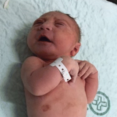


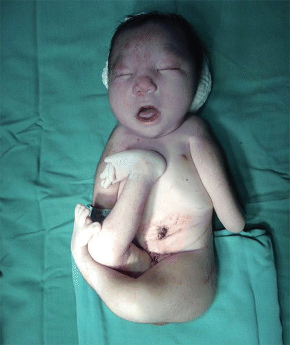
So far, it’s been linked to a 20-fold increase in microcephaly, in which newborns have irregularly small heads and underdeveloped brains.
Zika has also been tied to eye defects, hearing impairment and stunted growth.
In the case study, researchers looked at 438 patients from June 2015 to May 2016. About 17 of the fetuses came from moms who had documented Zika infection while another 28 had brain findings “suspicious” of the virus.


READ MORE: Here’s what Zika virus symptoms look like in pregnant women
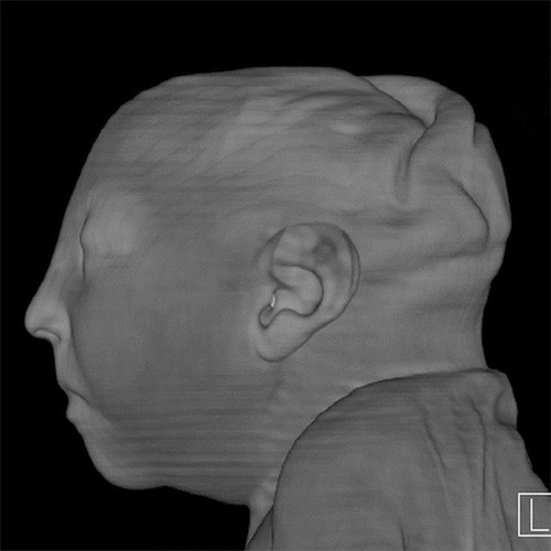
Turns out, a wide range of brain abnormalities cropped up, “including grey and white matter volume loss, brainstem abnormalities, calcifications, and a condition called ventriculomegaly, where the ventricles, or fluid filled spaces in the brain, are enlarged. Some babies infected by Zika may not have a small head size if the ventricles remain excessively enlarged.”
The skulls had ridges and protrusions in some cases, too.
“The unusual appearance of the skull, we hypothesize, is due to a combination of the small brain as it develops and a result of what, at some point, was likely a larger head size (due to ventriculomegaly) that then decompresses,” the researchers wrote.
See the full study and photos here.
READ MORE: What doctors know about how Zika virus potentially spreads
The latest Public Health Agency of Canada (PHAC) warning advises Canadian women who are expecting or hoping to get pregnant, to stay away from countries with Zika virus outbreaks, too.
As of Aug. 10, the World Health Organization identified 69 countries touched by the disease. They include much of South and Central America right into the Caribbean.
Canada has three maternal-to-fetal transmissions of Zika virus, including one case with severe neurological congenital anomalies. Another 220 travel-related cases have been documented, according to government updates.
Alberta alone has 26-lab confirmed cases of Zika. Twenty-five are from areas currently experiencing outbreaks – all were acquired due to travel.
READ MORE: Is Zika virus causing a spike in microcephaly in babies?
Zika is relatively harmless in adults, presenting with mild, flu-like symptoms in most people. Patients often encounter a headache, followed by a rash, lethargy and runny, red eyes as common symptoms.
carmen.chai@globalnews.ca
Follow @Carmen_Chai
Comments