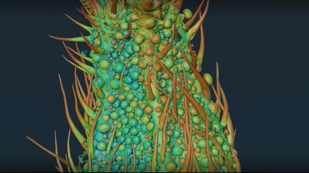
New highly detailed images taken by the Canadian Light Source in Saskatoon will help cannabis researchers to discover optimal harvesting times for the plant.

Teagen Quilichini is leading the research at the National Research Council and calls the images “groundbreaking.”
The research used the BXDS High Energy Wiggler Beamline and Mid-IR Beamline at the Canadian Light Source’s synchrotron to make highly detailed images of the plant and study its chemical composition.
“Due to the long prohibition, we have had very limited information and almost no research done on cannabis. It is time we catch up on that to inform the medical community and improve cultivation practices,” Quilichini said.
Quilichini’s team focused on the trichomes of the plant. These are the little sacs all over the plant that contain active compounds such as cannabidiol (CBD) and tetrahydrocannabinol (THC).

Get daily National news
“The trichomes are very sensitive and break easily, but by using the CLS beamline we were able to capture images that normal microscopes don’t even come close to. We can view all of the trichomes on the female flower, while still intact. That is groundbreaking in itself,” Quilichini explained.
The trichomes were analyzed during different phases in the plant’s development cycle. The research team discovered that the amount and types of trichomes vary heavily during the growth of the plant.
“This can inform better breeding and cultivation practices, but it will also allow us to select plants based on what we want them to do. It could greatly increase the chemical diversity of the cannabis plant and we could even discover new applications, instead of just trying to maximize THC.”
Cannabis research also helps further research on similar plants like lavender, mint and tomatoes.
- WestJet execs tried cramped seats on flight weeks before viral video sparked backlash
- Pizza wars? As U.S. chains fight for consumers, how things slice up here
- Health Canada says fake Viagra, Cialis likely sold in multiple Ontario cities
- Canada increased imports from the U.S. in October, StatCan says


Comments