
HALIFAX – A breakthrough in MRI technology developed in Halifax could soon help doctors better visualize complex surgeries before the first incision.

The Biomedical Transatlantic Imaging Centre, BIOTIC, recently received $2.9 million from the Atlantic Canada Opportunities Agency to develop software to create more detailed, accurate and comprehensive MRI images.
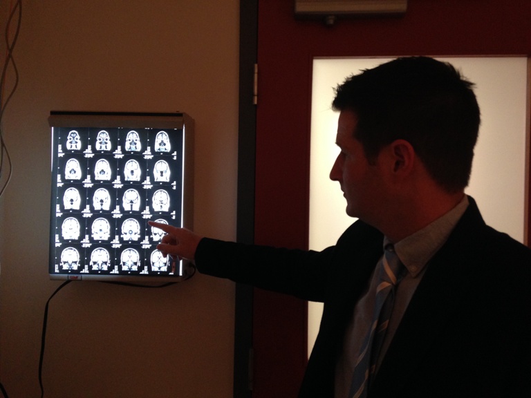
Dr. Steven Beyea, the scientific lead at BIOTIC, said the software will apply to three cases: patients with brain tumours, patients with prostate cancer and patients with liver disease.
Beyea said current MRI images can be challenging to read and difficult to process.
“If you come in and you’re about to have brain surgery, whether it’s for epilepsy or a brain tumour, a neurosurgeon is about to remove a portion of your brain. They, of course, want to understand exactly where critical brain functions are relative to that tissue they’re going to remove,” he said.
Beyea uses the example of a MRI scan done with the new software that shows doctors where a tumour is in relation to the part of the brain that controls the patient’s ability to move his or her hand.
“This information then is going to allow the surgeon to make better decisions about what to do even before surgery,” he said.

It’s something Kelly Marshall wishes was available when she had surgery to remove a benign brain tumour.


The Timberlea woman was diagnosed with the tumour about two years ago. But now post-surgery, she suffers from balance and sight issues as a result of fourth nerve palsy.
“It’s certainly something that happened within the brain from surgery so if they were able to have a little bit more detailed MRI imaging during or prior to maybe see something that maybe they couldn’t have seen, that would have been a huge help,” Marshall said.
Neuroradiologist Matthias Schmidt describes the technology as “the holy grail”.
“Information is going to be distilled into pictures that I can understand and information I can use to hopefully help patients have more accurate diagnosis, more confident diagnosis and that will hopefully lead to the most appropriate treatment,” he said.
“For the patient, this means they are so much more likely to make an excellent recovery and preserve the function that they have all the while getting rid of the tumour.”
In cases of liver disease, the software would map out what is liver iron and what is liver fat; in cases of prostate cancer, the software would help doctors determine where in the prostate gland the cancer is.
It is hard to tell whether the software could have helped in Marshall’s situation but she nevertheless said anything doctors can use to help patients like her is invaluable.
“If they have a tool that helps them with just one more fine detail, that is priceless to a patient who has a brain tumour.”
The funding from ACOA will be distributed over five years, at which time the goal is for researchers to produce software that can then go to market and be sold to hospitals worldwide.
Comments