


An international study out of London, Ont. is looking to see if a new type of imaging can better detect abnormalities in breast tissue.

The imaging is called digital breast tomosynthesis and is a type of 3D imaging. Researchers out of Lawson Research Institute want to learn whether it is better at finding abnormalities than the conventional digital 2D mammogram.
“When we only have two-dimensional views, it’s very difficult to separate a lesion from overlapping breast tissue, especially if the patient has dense breast tissue, which affects about 40 per cent of patients.”
During a tomosynthesis exam, the x-ray tube moves in an arc over the compressed breast and captures multiple images from different angles.
This is different from a conventional 2D mammogram, in which two X-ray images are taken of the breast: one from top to bottom, the other from side to side at an angle.

Get weekly health news
After the images are taken in the 3D exam, they are reconstructed into a set of 3D images by a computer which allows radiologists to examine the breast at multiple layers of depth, making it easier to distinguish normal breast tissue from potential abnormalities.

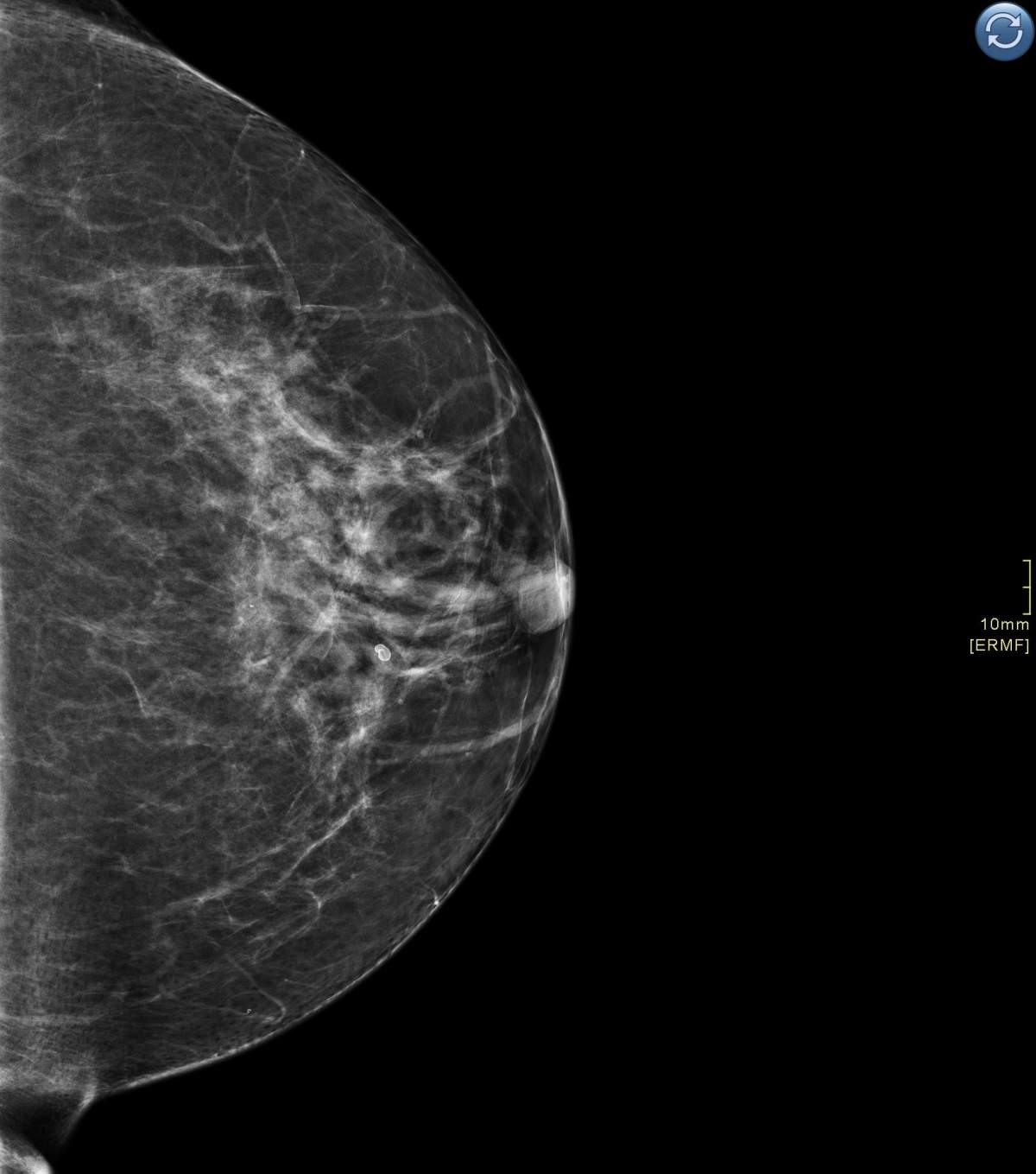

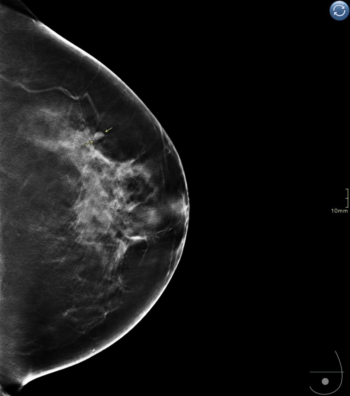


This is especially important for those with dense breast tissue because it may allow for screening at an early age than currently recommended, since before the 3D imaging, they weren’t able to see anything, she said.
The trial, called the Tomosynthesis Mammographic Imaging Screening Trial or TMIST, is happening at over 100 centres across Canada, the United States, and Argentina. Women are randomly selected to receive screening with standard 2D mammography, or digital 2D mammography along with the 3D imaging.
Over four years, the women will undergo either an annual or biennial screening with a long-term follow-up continuing for at least three more years.
The hope is the study will help radiologists evaluate whether the newer technology of 3D imaging is a more effective tool for detecting aggressive tumours.
Kornecki says the federal recommendation is to start getting mammograms at the age of 50 and to get one every two years. She says if a patient has a strong family history of breast cancer, they may be eligible to get a mammogram and MRI starting at the age of 30.

Through the Ontario Breast Screening Program or OBSP, women between the ages of 50 and 75 receive regular notices, encouraging them to schedule a mammogram for breast cancer screening.
Comments
Want to discuss? Please read our Commenting Policy first.